

Equine Diagnostic Imaging
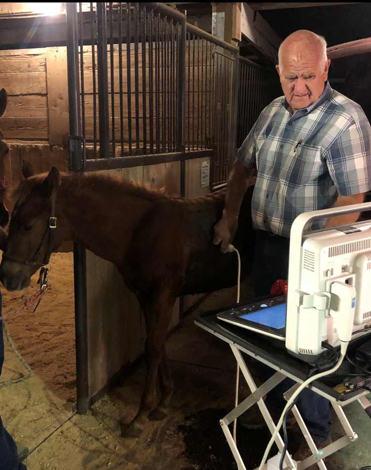
Equine Digital Ultrasound
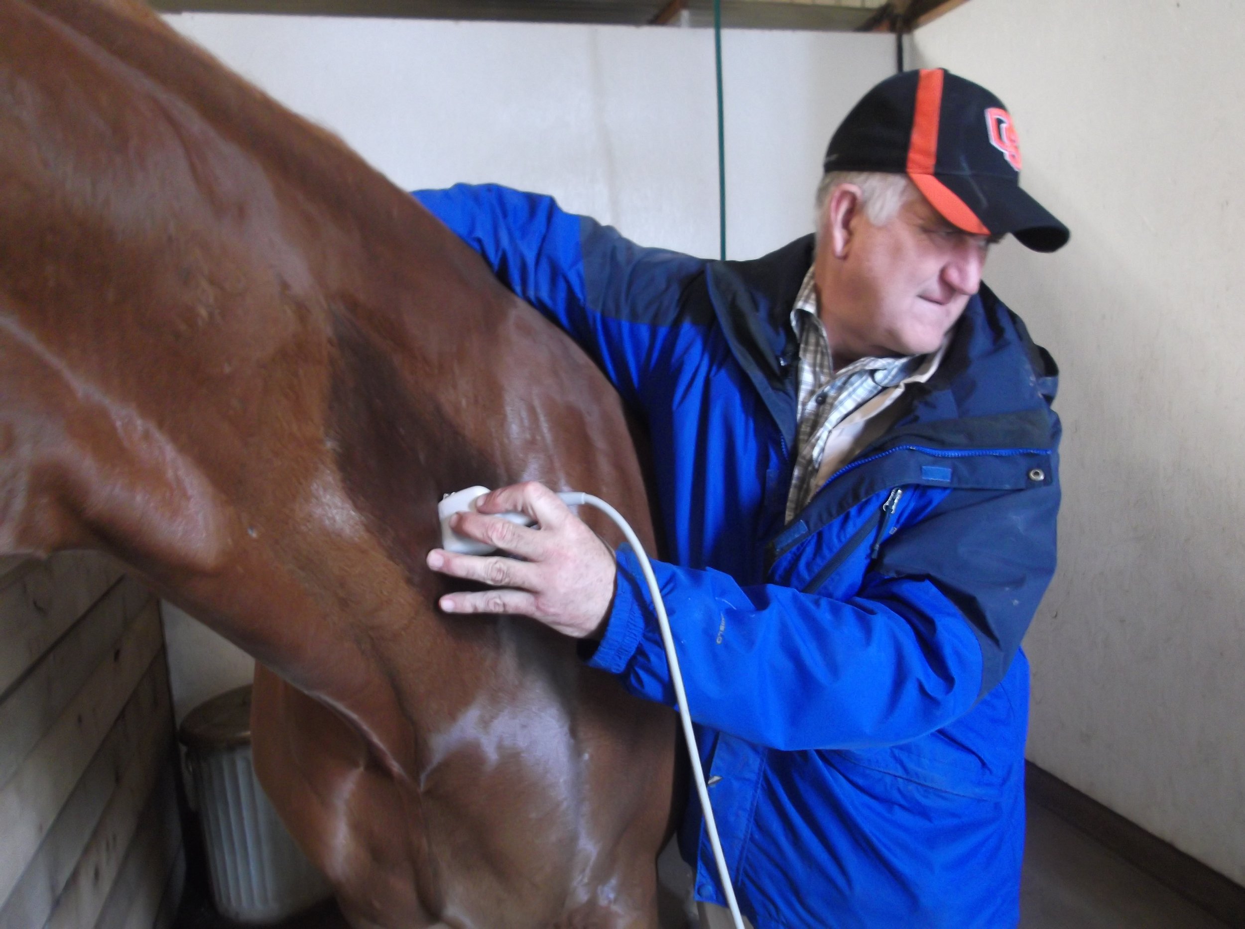
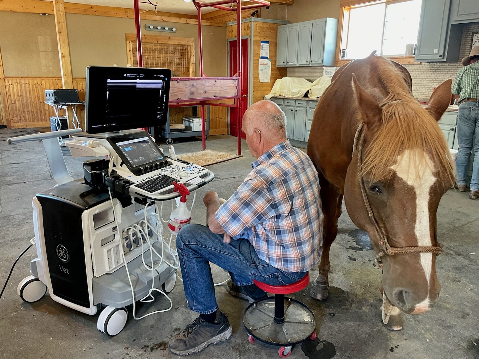
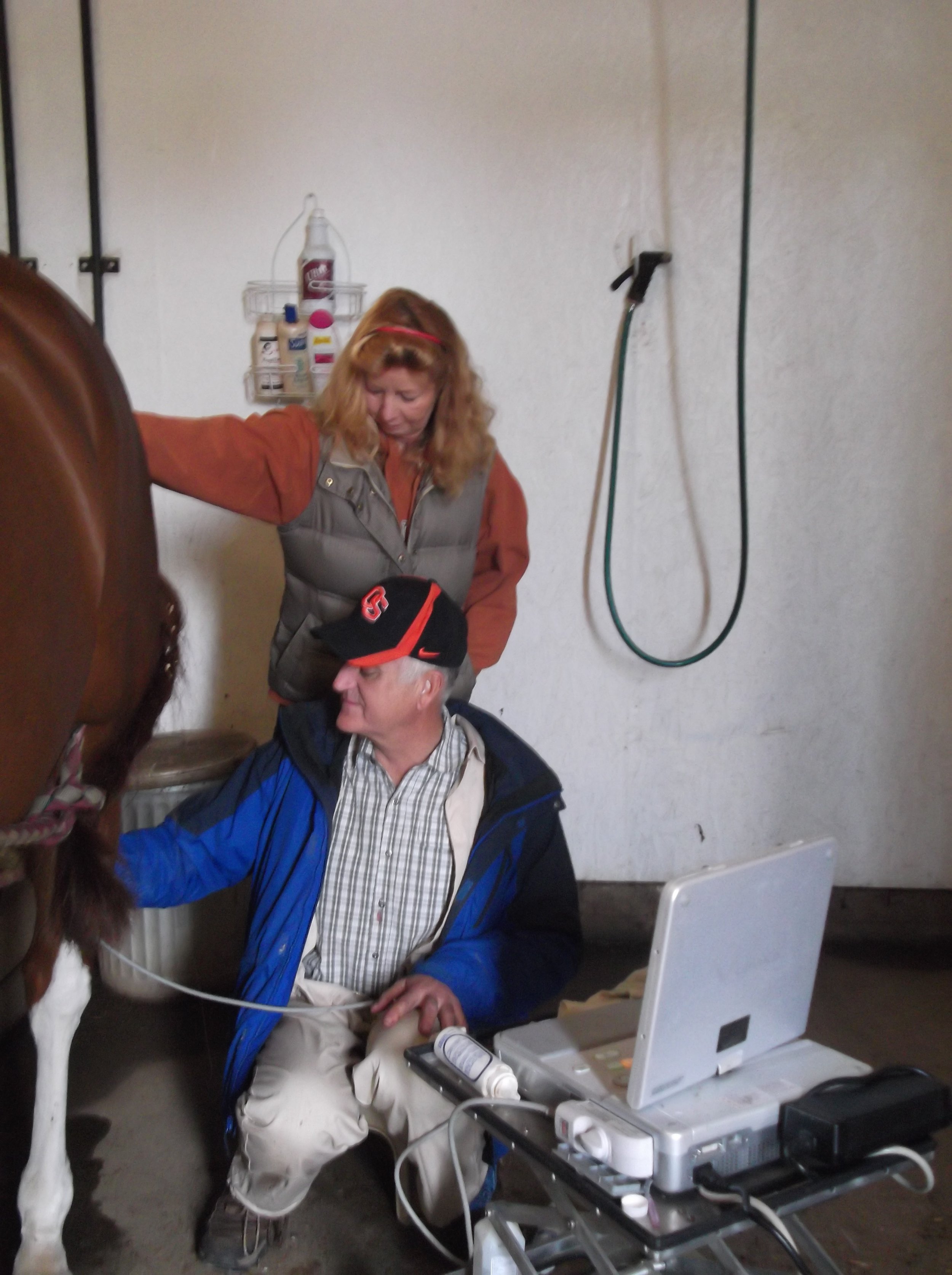
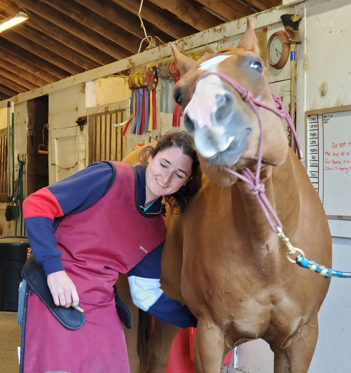
Equine digital ultrasound refers to the use of ultrasound technology to examine and diagnose conditions in horses. Ultrasound is a non-invasive imaging technique that uses high-frequency sound waves to create real-time images of internal structures.
In equine medicine, digital ultrasound is commonly used to evaluate various aspects of a horse's health, particularly in the musculoskeletal and reproductive systems. Here are a few applications of equine digital ultrasound:
Musculoskeletal Evaluation: Ultrasound can be used to assess soft tissue structures such as tendons, ligaments, and muscles. It can help identify injuries, tears, inflammation, or other abnormalities in these tissues. Equine athletes, particularly those involved in racing or jumping, often undergo ultrasound examinations to monitor the health of their tendons and ligaments.
Reproductive Assessment: Ultrasound is an essential tool for reproductive management in horses. It allows veterinarians to monitor the reproductive organs of mares, including the uterus and ovaries. It can be used to detect pregnancy, evaluate the stage of the estrous cycle, diagnose reproductive disorders, and guide the timing of artificial insemination or breeding.
Abdominal Examination: Ultrasound can be used to evaluate the abdomen and internal organs of horses. It can help identify conditions such as colic, evaluate the liver, spleen, and other abdominal structures, and assist in the diagnosis of gastrointestinal issues.
Cardiac Evaluation: Ultrasound can also be used to assess the equine heart. It allows veterinarians to visualize the structure and function of the heart, including the chambers, valves, and blood flow patterns. This can aid in the diagnosis of heart conditions and provide valuable information for treatment planning.
Digital ultrasound refers to the use of modern, digital imaging technology in the ultrasound machine. It allows for high-resolution images, improved image manipulation, and easier storage and sharing of ultrasound data.
Overall, equine digital ultrasound is a valuable diagnostic tool in veterinary medicine, providing veterinarians with detailed information about the internal structures of horses, helping them make accurate diagnoses and develop appropriate treatment plans.
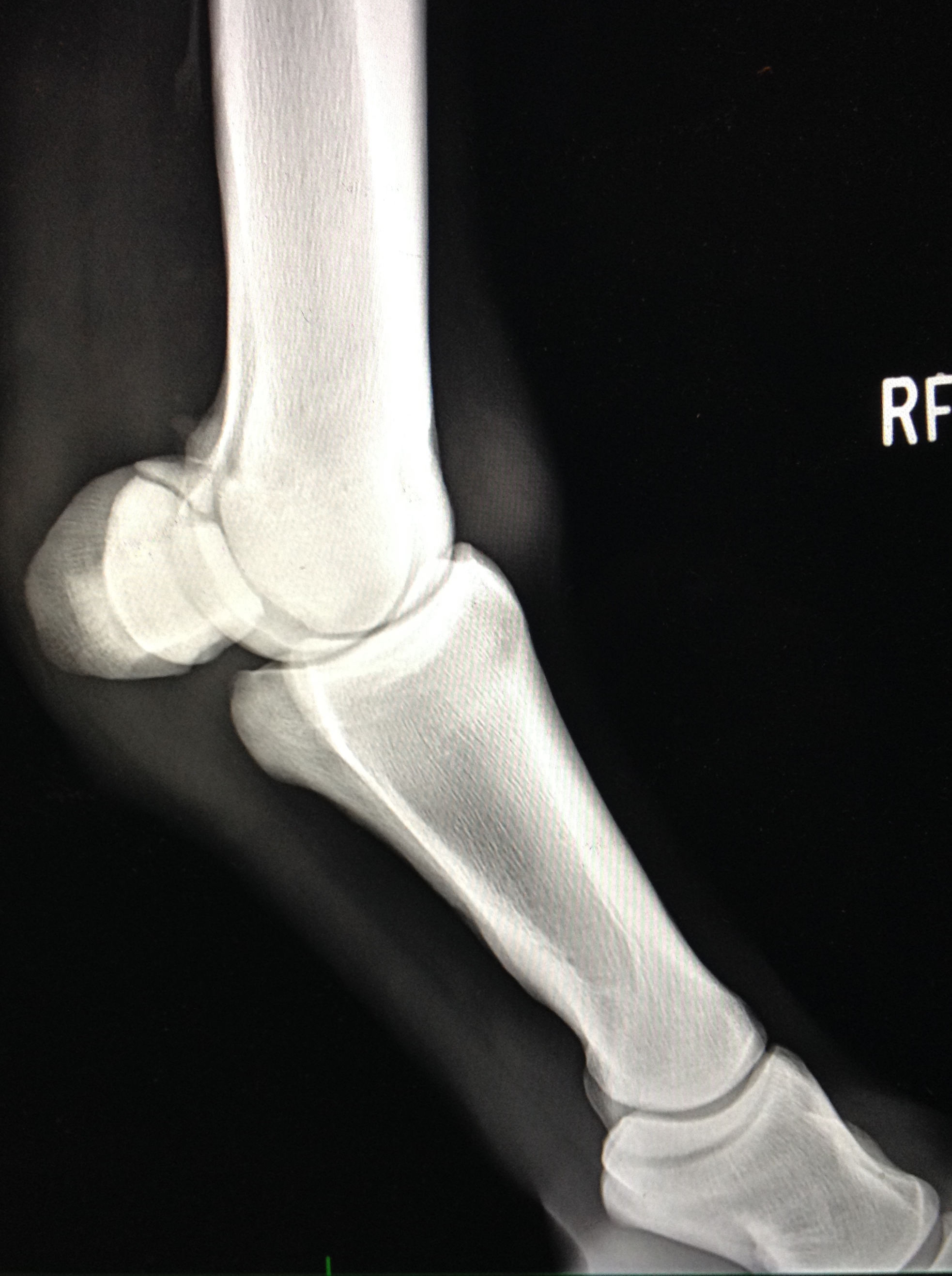
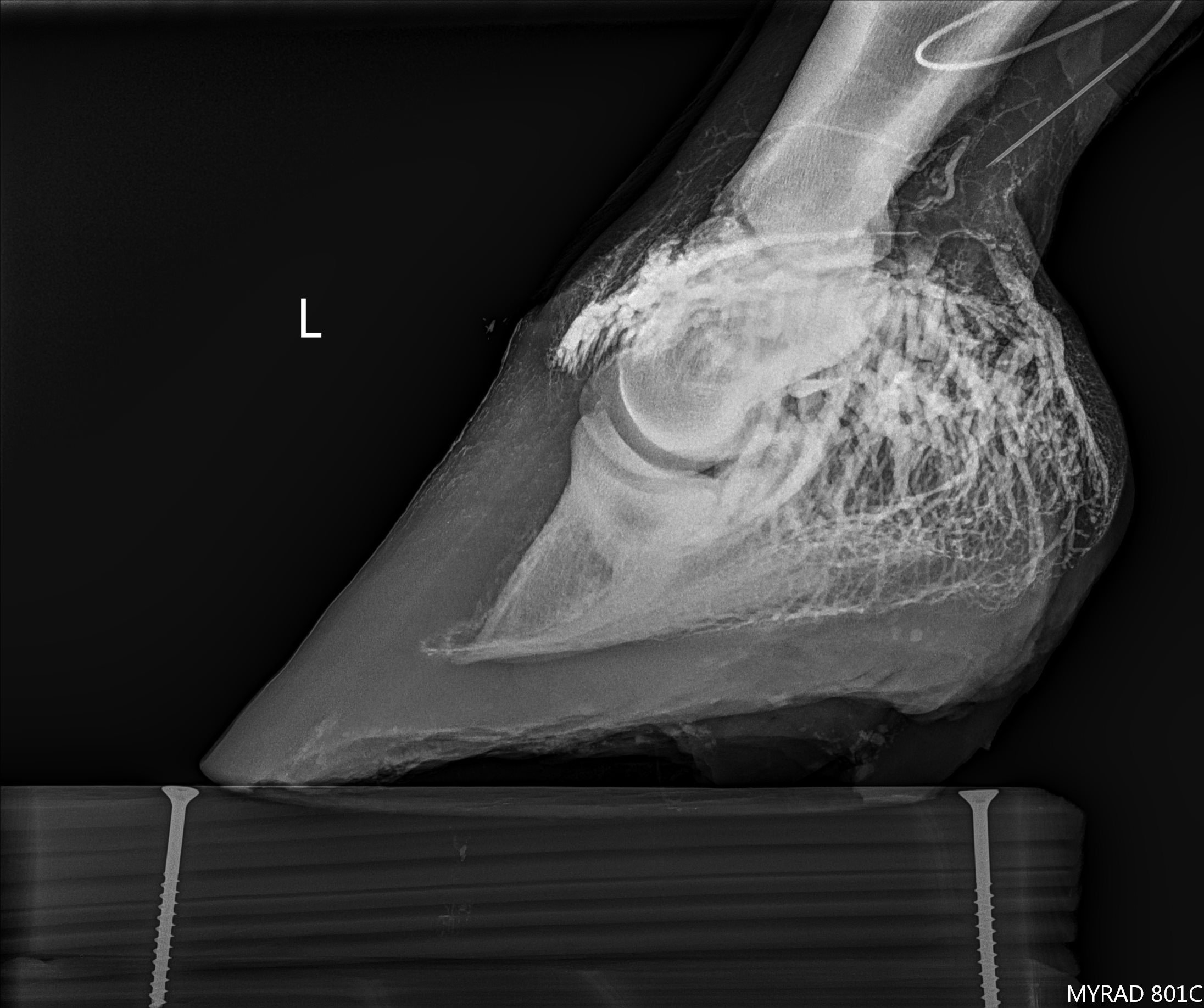
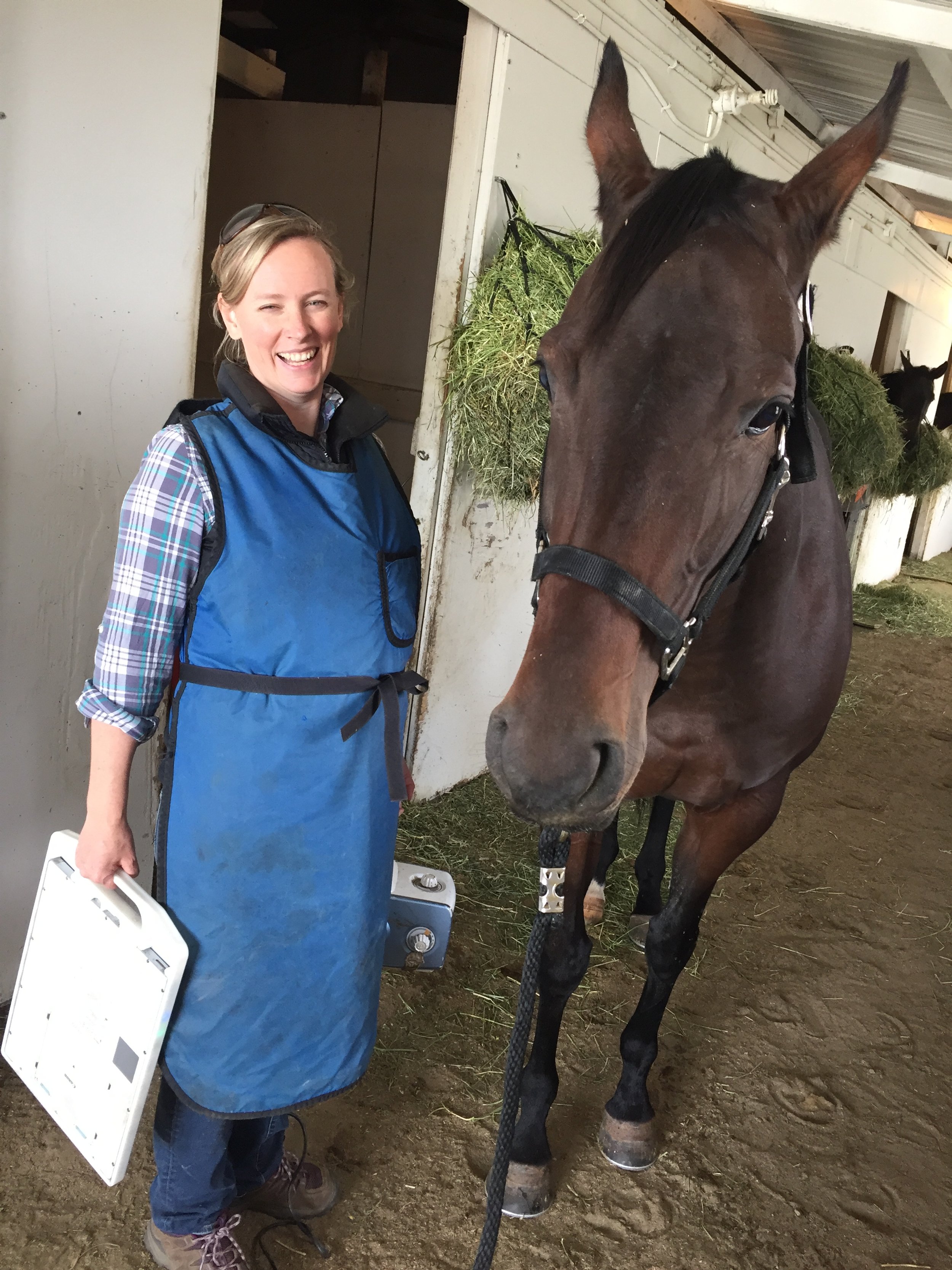
Digital radiography offers significant advantages over traditional radiography methods in evaluating joints, bones, hoof angles, and teeth. Here are some key benefits provided by a digital radiography (DR) system:
Consistent image quality: With digital radiography, the image quality remains consistent, reducing the chances of retakes due to poor exposure or image artifacts. This ensures that veterinarians have clear and accurate images for evaluation.
Adjustment capabilities: DR systems allow for adjustments in brightness, contrast, magnification, and edge enhancement. This flexibility enables veterinarians to optimize image visualization, making it easier to identify subtle bone details and evaluate soft tissue structures.
Increased efficiency: Digital radiography enhances efficiency by streamlining the image acquisition process. It reduces the time required per study, resulting in less sedation needed for the horse. This benefit is especially valuable for equine patients who may be sensitive to sedatives or require minimal stress during the examination.
Immediate image sharing: Digital radiographs can be quickly converted into JPEG format and emailed to referring veterinarians, allowing for instant communication and consultation. This prompt sharing of images facilitates collaboration among veterinary professionals, leading to timely diagnoses and treatment plans.
Portability: The portable nature of DR systems allows veterinarians to assess images on-site at horse shows, stables, or farms. This immediate evaluation capability enables efficient decision-making, especially in time-critical situations such as competitions or emergency farm calls.
Rapid fracture detection: Digital radiography helps in promptly ruling out fractures or catastrophic bone injuries. By providing instant access to high-quality images, veterinarians can make quick assessments, ensuring the safety and well-being of the horses.
Immediate results for pre-purchase exams: For pre-purchase exams, time is of the essence. With digital radiography, the results are available immediately, allowing potential buyers to make informed decisions about the horse's condition without delays.
Portable image storage: Digital radiographs can be easily stored electronically and accessed whenever needed. Owners or trainers can keep their horses' radiographs with them at all times, providing convenience and facilitating ongoing monitoring or consultation, even when they are away from the veterinary clinic.
Overall, the adoption of digital radiography in equine medicine at Oakhurst brings numerous advantages, including improved image quality, enhanced efficiency, immediate image sharing, and increased convenience for both veterinarians and horse owners.
Equine Digital Radiology
Equine Endoscopy/Gastroscopy
Oakhurst's endoscopy services provide valuable diagnostic capabilities for evaluating and visualizing various internal structures in horses. By using an endoscope and a large-screen monitor, veterinarians can examine the nasal cavity, sinuses, pharynx, guttural pouch, larynx, trachea, bronchi, esophagus, and stomach (gastroscopy). This allows them to identify potential issues that may be causing symptoms such as a bad attitude, poor performance, decreased appetite, poor hair coat, weight loss, or mild recurrent colic in your horse.
During the endoscopic examination, commonly known as "scoping," the horse is usually placed under light sedation or analgesia to ensure its comfort. This procedure enables the veterinarian to take biopsies and remove small foreign bodies if necessary. By visually inspecting the internal structures, the veterinarian can identify and diagnose various conditions or abnormalities that may be contributing to the horse's health issues.
If you notice any of the mentioned symptoms or suspect an internal problem in your horse, it is advisable to discuss the option of an endoscopic examination with your veterinarian. They will be able to evaluate the situation, determine if scoping is necessary, and provide appropriate treatment or management recommendations based on the findings.
Remember, early detection and intervention can often lead to better outcomes for your horse's health and well-being.

Call or email our office to bring your horse a day early if you would like us to help with the fasting procedure here on the farm!
503-554-0227 or office@oakhurstequine.com
AAEP explains more about Equine Gastric Ulcer Syndrome (EGUS) . . .
https://www.aaep.org/horsehealth/equine-gastric-ulcer-syndrome
Consultation Services
Dr. Jack Root provides expert equine consultation services with a focus on pre-purchase clinical exam reviews, lameness evaluations, and radiograph interpretation. While not a board-certified radiologist, Dr. Root brings over 45 years of hands-on experience in equine sports medicine and lameness—ranging from Thoroughbred racehorses to English sport horses, Western performance horses, and beloved backyard companions. He has extensive training in radiology and diagnostic imaging, which he applies daily in his work. As a member of the International Society of Equine Locomotor Pathology (ISELP), Dr. Root remains committed to advancing his knowledge and staying at the forefront of equine lameness diagnostics. His lifelong dedication to continuing education and excellence in imaging has shaped the distinguished career he leads today.
Pre-Purchase Exam Consultation
For both local and out-of-town clients seeking to acquire horses, Dr. Root’s expertise comes to the fore. By diligently examining existing clinical reports and digital imaging materials via email, he provides impartial and informative assessments. These evaluations empower clients to make well-informed decisions when considering a horse purchase.
Lameness Consultation
Meticulous lameness examinations encompass a multifaceted approach. This includes assimilating owner history, observing the horse in motion, conducting musculoskeletal palpation and manipulation, administering diagnostic nerve blocks, and, when required, employing advanced imaging techniques such as Digital Radiography, Digital Ultrasonography, Nuclear Scintigraphy, and MRI. This comprehensive methodology is designed to pinpoint the root cause of pain or diminished performance in equine athletes.
Occasionally, obtaining a second opinion from another Equine Veterinarian can prove invaluable with a challenging lameness case.
For inquiries or to schedule a consultation, please direct your correspondence to office@oakhurstequine.com. Or call our office at 503-554-0227. Pricing is tailored to the volume of images and the duration of consultations.