
When a Hematoma Turns to Stone: Dystrophic Mineralization in Horses
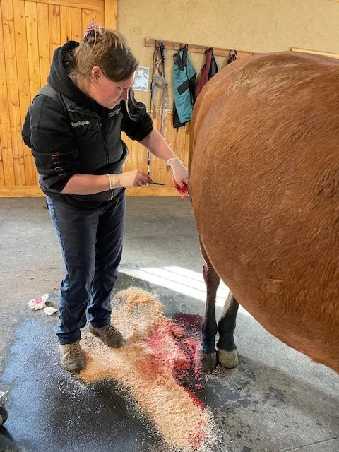
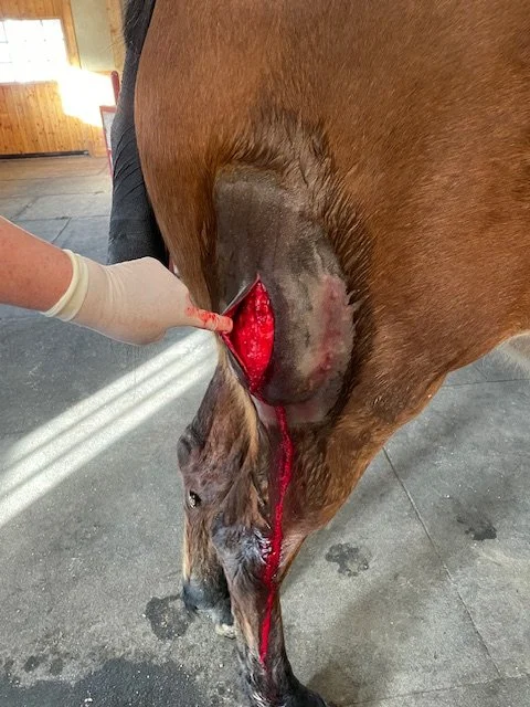
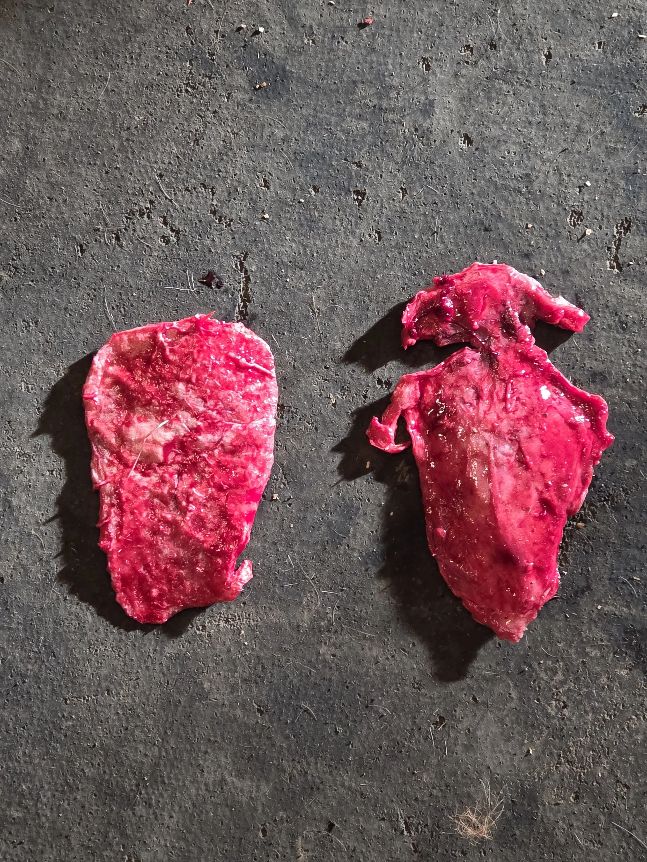
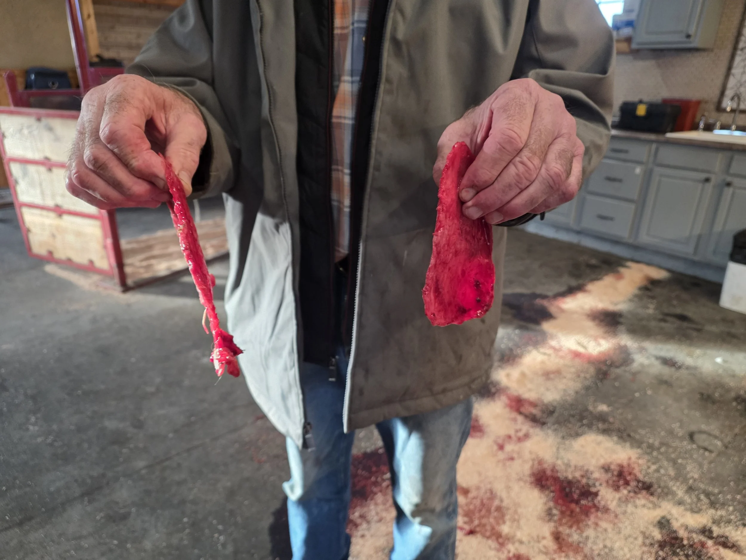
At Oakhurst Equine Veterinary Services, Dr. Flynn Magorian recently managed a fascinating and educational case that earned high praise from Dr. Root, involving the removal of two large calcified plaques—one surprisingly overlying the other. While large hematomas and seromas are fairly common in horses following blunt trauma, kicks, or even routine surgical procedures, they usually resolve with time and appropriate care. Occasionally, though, healing doesn’t follow the usual script. In rare cases, the body lays down hard, eggshell-like mineralized tissue beneath the skin, a process known as dystrophic mineralization of an organized hematoma. Dramatic in appearance but localized and non-systemic, this condition highlights the importance of understanding not just how horses heal—but how and why healing can sometimes take an unexpected turn.
What Is Dystrophic Mineralization?
Dystrophic mineralization occurs when calcium salts and other minerals are deposited into damaged or necrotic tissue, even though blood calcium and phosphorus levels are completely normal. In horses, this most often develops following:
- A large hematoma or seroma/recurrent ones
- Inadequate drainage
- Repeated motion or trauma at the site
- Chronic inflammation during healing
Over time, the body organizes the blood clot or fluid pocket with fibrous tissue. Changes in local tissue chemistry then allow calcium phosphate to precipitate, forming rigid, laminated plates or shells. On palpation, these can feel strikingly similar to thick eggshells or rock-hard armor beneath the skin.
What Does It Look and Feel Like? Clinically, these lesions are usually:
- Firm to rock-hard
- Subcutaneous (beneath the skin)
- Sometimes considerable surface area
- Often non-painful once mature, but may be annoying when manipulated
- Poorly compressible
- Sometimes irregular or plate-like in shape
The overlying skin may appear normal, although chronic pressure can lead to thinning, reduced mobility, or—in rare cases—ulceration. Common locations include the shoulder, hip, thigh, neck, and chest wall—areas prone to trauma and movement. A five-year-old horse is a very typical age for this condition.
How Is It Diagnosed?
Radiographs X-rays are usually diagnostic and reveal:
- A mineralized outer shell
- Sometimes a more lucent center if residual fluid remains
- Ultrasound
- Ultrasound can help determine:
- Thickness and surface area of the lesion
- Presence of remaining fluid
- Involvement of surrounding soft tissues
Bloodwork: Serum calcium and phosphorus levels are typically normal, reinforcing that this is a local—non systemic metabolic—process.
Conditions That Can Look Similar Several other conditions may enter the differential diagnosis, including:
- Myositis ossificans (true bone formation within muscle)
- Chronic abscess with mineralized capsule
- Foreign body granuloma
- Soft tissue tumors with dystrophic calcification
Imaging usually allows these to be distinguished without biopsy.
Does it need to be treated?
Not always.
Conservative Management
If the lesion is:
- Stable
- Non-painful
- Not interfering with tack, movement, or performance
It can often be left alone and simply monitored. Once mineralized, these lesions rarely regress, but many remain clinically insignificant. Surgical Removal Surgery is considered when the mass:
- Interferes with athletic function or tack
- Continues to enlarge
- Causes skin compromise
- Is a cosmetic concern in sale horses
Complete excision is typically curative, though surgery requires careful technique. These mineralized plates may fracture during removal and often leave significant dead space, making drains and compression bandaging important for successful healing.
Prognosis
The prognosis is excellent. This condition does not affect overall health or longevity, and athletic performance is usually unaffected unless the lesion interferes mechanically. Recurrence is uncommon when the mineralized tissue is fully removed.
Take-Home Message
An “eggshell-like” mass forming under the skin months after a large hematoma can be alarming—but it is usually a benign, localized result of the body’s healing process. With proper diagnosis and thoughtful management, dystrophic mineralization is more of a curiosity than a catastrophe.
If you notice a hard mass developing where your horse once had a significant bruise or swelling, give Oakhurst Equine Veterinary Services a call for a consultation. In most cases, the news is far better than the texture suggests.